
Research materials
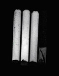
Foam phantomsTest objects were manufactured in Medical Physics Department, University of Dundee, Scotland, in the form of glass tubes shown in Fig. 1. Three of them contained synthetic foams of different porosity, the fourth one contained glass beads. The tubes were filled with a special agarose gel providing relatively long T2 relaxation time. The images were acquired using Siemens Magnetom 1.5T scanner in German Cancer Research Centre (GCRC), Heidelberg.
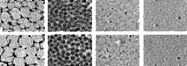
The images showing cross-sections of tubes filled with foams were acquired at FOV=100mm x 100mm and FOV=200mm x 200mm, with the image matrix size equal to 256x256 pixels. Fig. 3 presents magnified phantom images (starting from left side):
Slice thickness is equal to 4mm for upper-row images and 2mm for lower-row images, respectively.
<< Back | Optical foam images >> |