
Research materials
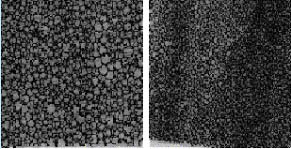
Optical foam imagesIndependently, to compare with MRI images, the optical foam images were investigated. Foam cross-sections images were obtained using optical scanner. The images were scanned for high porosity foam (shown on the left in Fig 4) and for low porosity foam (shown on the right in Fig. 4).
<< Back | Polystyrene phantoms >> |